Hi everyone,
I would like to know if it is possible to use 5ttgen or any similar without a T1 image as I don’t have any available for my data (only diffusion and T2) ?
Thanks for your help
Céline
Hi everyone,
I would like to know if it is possible to use 5ttgen or any similar without a T1 image as I don’t have any available for my data (only diffusion and T2) ?
Thanks for your help
Céline
Hey celinede,
you may check out this Info which I found at another post.
dwi2response dhollander is an alternative, often much more accurate, way of obtaining multi-tissue (WM, GM, CSF) response functions from your data. It doesn’t need a co-registered T1w image, or 5tt segmentation thereof. For more information on the algorithm, see this abstract. The method only needs your (preprocessed) DWI data, and automates all other steps to obtain the response functions.
Best
Max
Hi Céline,
While the existing 5ttgen algorithms are designed to operate on T1-weighted images, that’s not to say that it’s impossible to produce a 5TT image unless you have a T1-weighted image; only that a script to automate the generation of such has not yet been written / provided. So perhaps an alternative question would be: Is it possible to perform reasonable tissue segmentation using only a T2-weighted image? If so, it’s possible to convert the output from said segmentation into the 5TT format, and it would additionally be possible to automate that process in a script.
dwi2response dhollanderis an alternative, often much more accurate, way of obtaining multi-tissue (WM, GM, CSF) response functions from your data.
While this will give multi-tissue response functions, what is ultimately needed in terms of generating a 5TT image is a multi-tissue decomposition in every voxel; this is provided using the dwi2fod msmt_csd algorithm (for which dwi2response is typically a prerequisite). But one way a 5TT image can be used is in the dwi2response msmt_5tt algorithm; hence the confusion.
I think we’ve all thought about using such decompositions from MSMT-CSD as inputs to ACT, but I don’t know if anybody’s actually done the experiment. A potential disadvantage of defining the 5TT image based on the diffusion data is that you don’t get to exploit the elevated spatial resolution of an anatomical contrast image; but then again, tissue segmentation based on a single 3D image can be erroneous in other ways…
Dear all,
Sorry for bringing this up! Looks like it’s been a while since the last post. I am facing to a similar situation where I only have T2-weighted to perform tissue segmetation in MRtrix.
I have been recently started working on MRtrix and currently facing the same issue. In fact, I have got FOD image from DWI using dwi2response tournier algorithm. Now I want to apply ACT method which requires me to have 5tt segmented image. However, I only have T2-weighted image.
I found this post telling that in order to perform tissue segmentation using only T2-weighted image, dwi2response dhollander is an alternative. I have some related questions which hope to receive your suggestions:
Thank you very much,
Best,
Van
I found this post telling that in order to perform tissue segmentation using only T2-weighted image, dwi2response dhollander is an alternative.
This is not a correct interpretation of that text. dwi2response dhollander is a DWI response function estimation algorithm. While a DWI b=0 image is largely T2-weighted, and that particular algorithm does involve some degree of tissue segmentation in the process of deriving those response functions, it requires more than just T2-weighted data (ie. it requires b>0 images also), and it should not be considered “a tissue segmentation algorithm”.
I would instead repeat myself from the earlier post: is it possible to perform tissue segmentation using only a T2-weighted image, using any software? If such segmentation can be achieved, then producing a 5TT image for ACT from that segmentation is simply a matter of format conversion.
Hi Smith,
Thank you so much for your clarification.
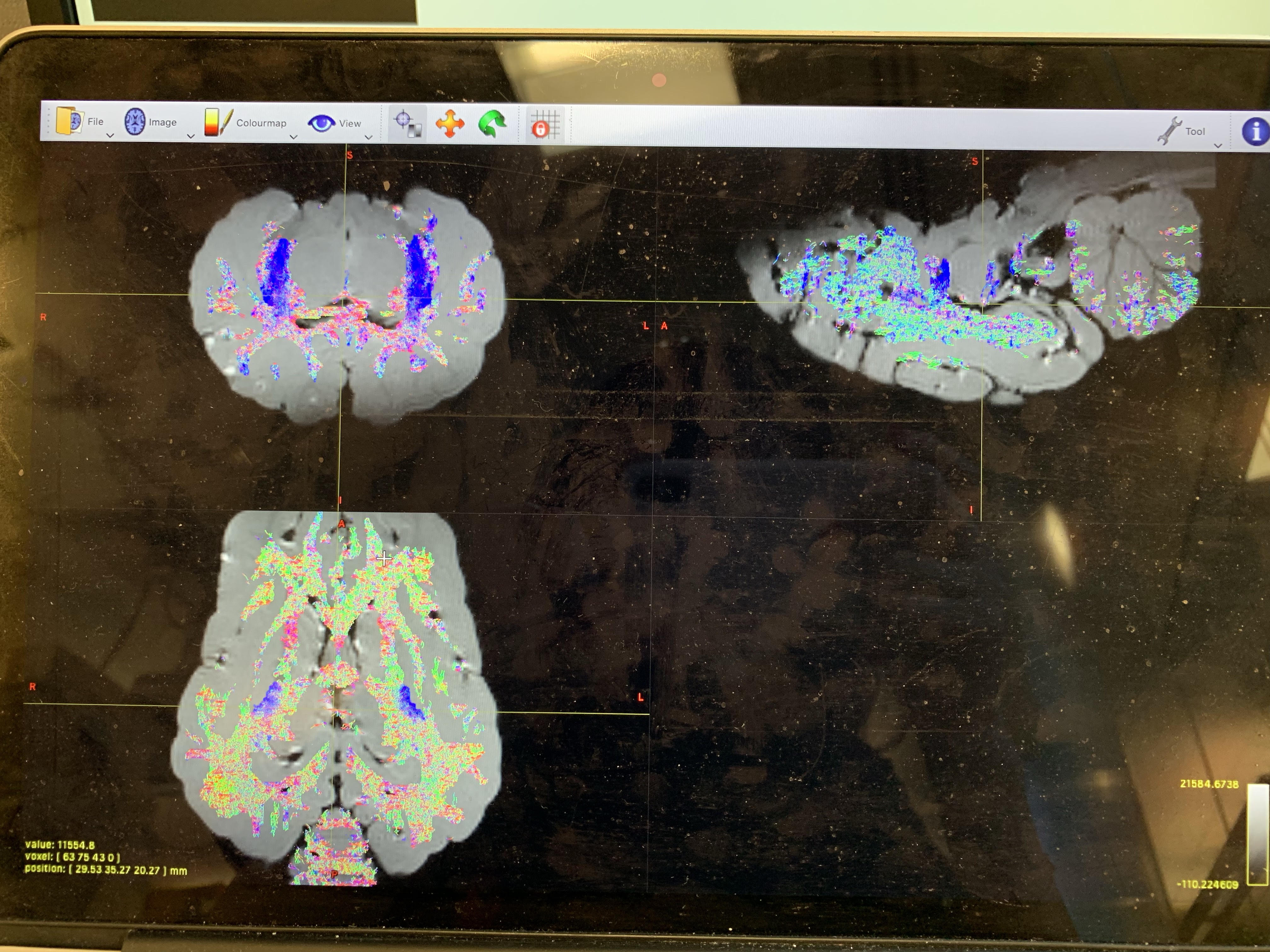
I have been using dwi2reponse dhollander to create 3 response text files and 3 FODs of WM, GM and CSF. In fact, the scans that I am using are ex-vivo scans. Instead of using ACT algorithm, I have tried using tckgen with an option -seed_image. The seeding image was obtained by dwi2response dhollander. Details are described below.
After I received WM FOD image, I assumed that FODs regions represent for WM regions. Thus, I applied threshold on FOD WM image to get out the region of WM only, named as WM_threshold.mif. After that, I fed WM_threshold.mif to a command of tckegen. It looks like below:
tckgen WM_threshold.mif -seed_image WM_threshold.mif -select 100k track.tck
I received a track files where most fibers are running within WM regions. Even though the results are not so perfect, I found it acceptable still. Could you please help me to verify this method?
I attached an overlay DWI-tractography loaded here for a reference.
Many thanks,
Jane
I have been using dwi2reponse dhollander to create 3 response text files and 3 FODs of WM, GM and CSF.
This description is incorrect, which is the source of confusion. dwi2response is only responsible for response function estimation; FOD estimation is achieved by the dwi2fod command.
After I received WM FOD image, I assumed that FODs regions represent for WM regions. Thus, I applied threshold on FOD WM image to get out the region of WM only, named as WM_threshold.mif. After that, I fed WM_threshold.mif to a command of tckgen.
Generating a binary brain / WM mask for seeding / tracking is an entirely common step.
Even though the results are not so perfect, I found it acceptable still. Could you please help me to verify this method?
It’s difficult to verify from a single image, but there seems to be less colour coherence over distance and more kind of “speckly” colours. I’d be looking more closely at the underlying FODs, just to be sure that the orientations are coherent, and ideally also that they are pointing in the right direction with respect to the underlying anatomy.
Thank you very much Rob!
I will have my collaborator double check on fiber orientation anatomically.
Best,
Jane