I am using the ISMRM tutorial for generating the fiber tracts. It is working fine for me. I am trying to introduce some breaks to the fiber tract by simulating some hole like structures (just like ventricles) inside the white matter. For achieving this, I am trying to copy the values from ventricles and paste at desired locations in the white matter. I am not quite sure about which files I need to edit? Any idea?sample.PNG|690x337
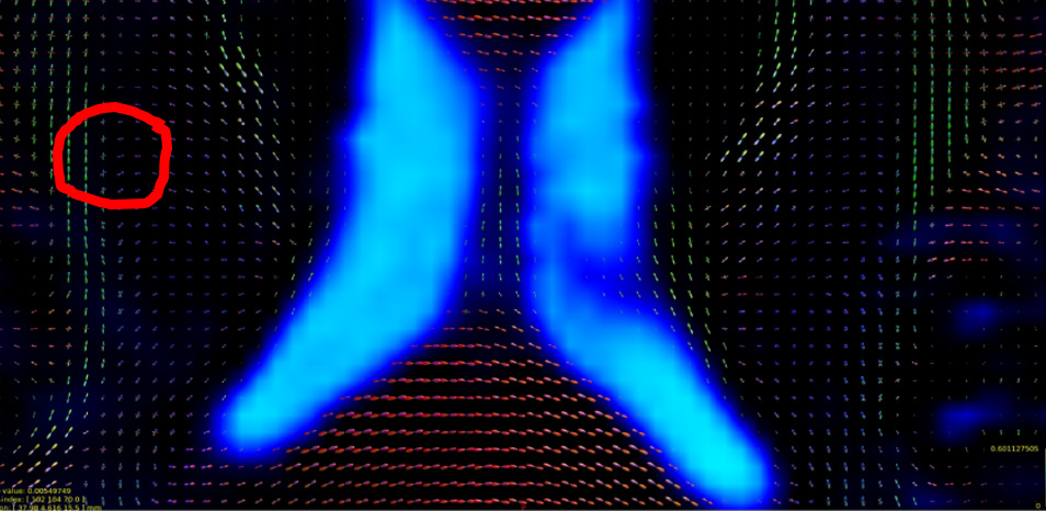
In the attached image you can see that there are no DTI direction glyphs inside the ventricle(blue region) because it if fluid filled. Is it possible me to create a small artificial fluid filled location(inside red circle) in the white matter by simply copying things from ventricles and pasting in the white matter areas? If that is possible, which files I will have to edit?
Welcome @reshma!
Firstly I would maybe propose a clarification on your topic title. Given the range of data with which I deal, I typically use the phrase “connectome” to refer specifically to the matrix of inter-areal connectivities that results from assigning streamlines from the tractogram to nodes of a parcellation. The reason I think that particular definition is important here is because, within the general domain in which you’re working, which we’ll call “simulated pathology”, it’s possible to perform manipulation of the data at multiple locations along the reconstruction pipeline; one of those locations is within the connectome matrix data itself, modulating the values stored within individual entries; but that is not what you’re wanting to do here, despite the title possibly hinting in that direction.
So ultimately what you’re looking for is a way to introduce simulated pathology into image data, that will influence the streamlines tractography reconstruction. I would then break this into three possibilities:
-
Influence the DWI data (whether it be the actual raw diffusion-weighted data, or the parameters of a diffusion model);
-
Influence the anatomical segmentation so that ACT applies different priors;
-
Use manual regions of interest to prevent streamlines from traversing a given region.
The precise behaviour of the tractography algorithm in the presence of such modifications may vary slightly depending on which approach you use and how.
As far as how to actually perform such manipulations, take a look at the following commands:
-
mreditcan generate a ROI image containing a sphere at a given location with a given radius; -
5tteditis tailored for modifying ACT 5TT images; -
mrcalcutilising a multiplication operation can modulate the values stored within an image at given locations, as long as you can provide an appropriate image with which to perform that modulation (e.g. multiplying by 0 within the region and 1 outside the region would null the image within the region but leave all other values unaffected).
Hope that’s enough to get you started
Rob
Thank you Robert for your reply. Yes, I am looking for a way to do simulated pathology. Because of some pathologies, the white matter region in the brain may have fluid filled holes just like the ventricles. I already managed to prevent connectome generation by manually editing the nodif_brain_mask.mif file. Thanks for your inputs, now I can think of trying those options as well. Also, thanks a lot for your time elaborating on this. Cheers!