I made a standard calulation 3 x b-vallues with the “dwi2response dhollander” and the multitissue “dwi2fod” and the WM FOD file gives me FODs in the CSF and the GM FOD file gives me just spheres. The CSF file also has only spheres.
This is the expected behavior of the algorithm. The GM and CSF are modeled as isotropic compartments. Probably the one you are interested in is the WM FOD.
Non-zero WM FODs within CSF is entirely possible. There’s no “guarantee” that doing multi-tissue CSD will completely nullify such. It depends on the response functions and your data. E.g. If your SNR is very low, and you have b>0 volumes where the intensity in CSF is not perfectly equivalent between volumes, the algorithm may use the anisotropic nature of the WM response function to “explain” some of that variance, even if that RF is not a perfect match for the signal decay as a function of b-value.
GM FOD multi tissue gives me just spheres.
The automatic determination of lmax for output ODF images is based on the lmax of the input response functions. Since dwi2response dhollander is explicitly coded to yield isotropic response functions for GM and CSF, using those RFS for deconvolution can only yield lmax=0 ODFs, i.e. spheres.
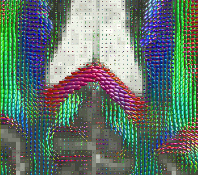
But I’m also curious to know that the background image is in your screenshot. It looks highly discretized like a fixel count image, but the contrast looks like a b=0. Asking in case there’s some fundamental problem with your upstream processing.