Hi all MRtrix experts,
Are there any methods that can reduce the false-positive FOD lobes when voxels are near the bone or air regions?
Thanks!
Regards,
John
Hi all MRtrix experts,
Are there any methods that can reduce the false-positive FOD lobes when voxels are near the bone or air regions?
Thanks!
Regards,
John
Dear John,
Interesting question! To diagnose your problem, we need much more information:
What data are you using? (b-values, no. of directions, and spatial resolution)
How do you currently compute your FODs? (including preprocessing)
Screenshots of the erroneous FODs
Cheers,
Ben
Hi Ben,
Thanks for your reply!
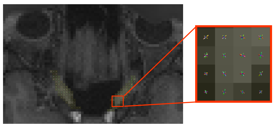
I used HCP data (multi-shell data with b=1000/2000/3000 s/mm2, 288 dirs, and 1.25×1.25×1.25mm³ voxel size) to reconstruct FOD in the optic nerve region.
The data have already been well preprocessed, so I directly generate the FOD using msmt-csd algorithm.
Here is the visualization of the FODs in the optic nerve.
Hi John,
Having the outcome of the analysis not meet your expectations requires assessing the quality of every step along the way; it’s unfortunately not as simple as flicking a switch to improve FOD quality.
I’ve myself recently experimented with trade-offs between spatial resolution and signal, and while higher spatial resolution looks quite nice at the cortex, it can lead to noisy data in non-peripheral regions where no individual coil has very good sensitivity. So you’d want to convince yourself first that the quality of the HCP data in this region is actually OK. In addition to e.g. dwishellmath mean, you could also look at the raw DWI data plotted on the sphere (“Load dixel ODF” in mrview ODF toolbar) to see if the shape of the raw data makes sense with respect to the anatomy.
Beyond that, given the fact that you’re using pre-processed data, the key thing to check would be your response functions; where were they generated from, what do they look like relative to one another. Do the FODs look OK elsewhere in the image, but no good in this specific pathway (which would circle back to my first point)?
Rob