Hi Community,
I ran the following command:
dwi2fod msmt_csd DWI.mif RF_WM.txt WM_FODs.mif RF_GM.txt GM.mif RF_CSF.txt CSF.mif -mask nodif_brain_mask.nii.gz
There is definitely something odd-looking about that corpus callosum, particularly in anterior part where it lines the ventricles - the rest of the brain looks good. I doubt it’s anything to do with the CSD itself (I expect you’d find similar issues using a tensor-based analysis - easy enough to verify if you need to).
I recommend you look through your raw data and check whether any of your volumes are corrupted in this region.
Also, maybe you can tell us a bit more about your acquisition parameters (b-value, number of directions, resolution, etc), and what processing you’ve done on these data/
How to judge whether the results of the dwi2fod is right?
Please give me some advice, thank you!
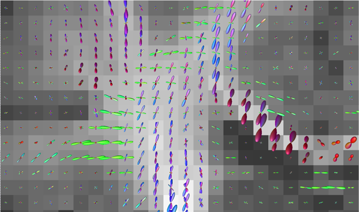
More generally, if you’re trying to do a visual assessment of FOD estimation in a healthy brain, what you’re looking for is: Estimated fibre orientations that are or are not consistent with the spatial continuity of the bundle.
For instance, take a look at this zoomed coronal view of the centrum semiovale where the FODs are clearly “wrong” (looks like pathways with high fibre density just vanish):