Hi, Rob,
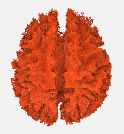
Having said that: Are you sure that the top image is the one without pre-processing steps? The bottom one looks cleaner and the brain is more conventionally shaped :-/
I’m pretty sure that Fig11 is the one without pre-processing steps. I just want to see what the figures will be if I discard the “dinoise”, “preproc”, and “bias-correct” steps, because these three steps take a lot of time. However, the results seems quite cool and unbelievalbe.
Also, as Donald said this data may not suitable for pre-processing ,as they are not reversed phase-encoded EPI images.
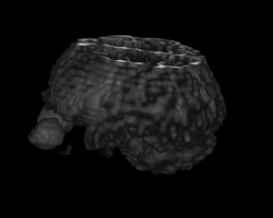
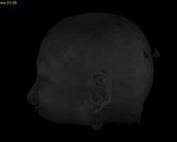
I’m sorry I should indicate in the post that Fig 10 and Fig 11 are the results of different subjects. The data is problematic, as shown in re-edited figures. They have been cut-off which may have a great influence on registration from T1 image to DWI image, especially the data of Fig 10.
As I think, It explains why Fig 11 looks cleaner and the brain is more conventionally shaped.
I don’t know why ACT works well with unpreprocessed Single-Shell DWI data. Anyway, it seems works.![]()
Also, I’m curious about the reason for this phenomenon.
By the way, does ACT work with Multi-Shell DWI data?
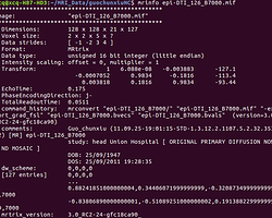
I’ve test on a problematic Multi-Shell data, as Fig 12 shows. It seems also works.
Fig 12:(Multi-Shell DWI data)
Problematic Datasets… ![]()
![]()
![]()
Many Thanks,
Chaoqing


