Hello,
I am running 5ttgen fsl using a brain-extracted T1 image that shows relatively low contrast in the circled area:
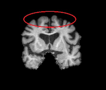
I used 5ttgen fsl T1_brain.nii.gz 5TT.nii.gz -premasked -nocrop
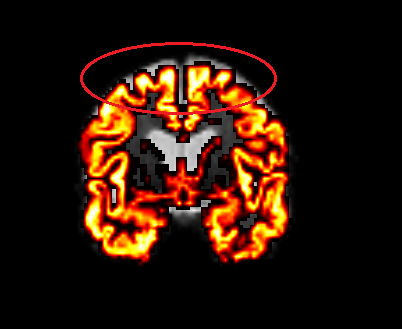
The overall segmentation in T1 space as well as after registration to DWI space looks good, however, in the circled region, grey matter appears overestimated:
In the ACT-based tractography streamlines seem to terminate incorrectly in that region:
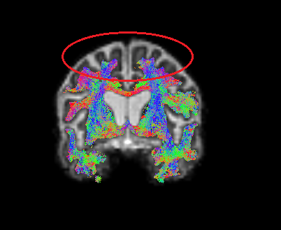
This issue is consistently present in subjects from one study center but is not observed in data from other centers processed with the same pipeline. Could this be related to reduced T1 contrast in that region? Do you have suggestions on how to improve the segmentation in such cases?
Thank you very much for your help,
Franziska