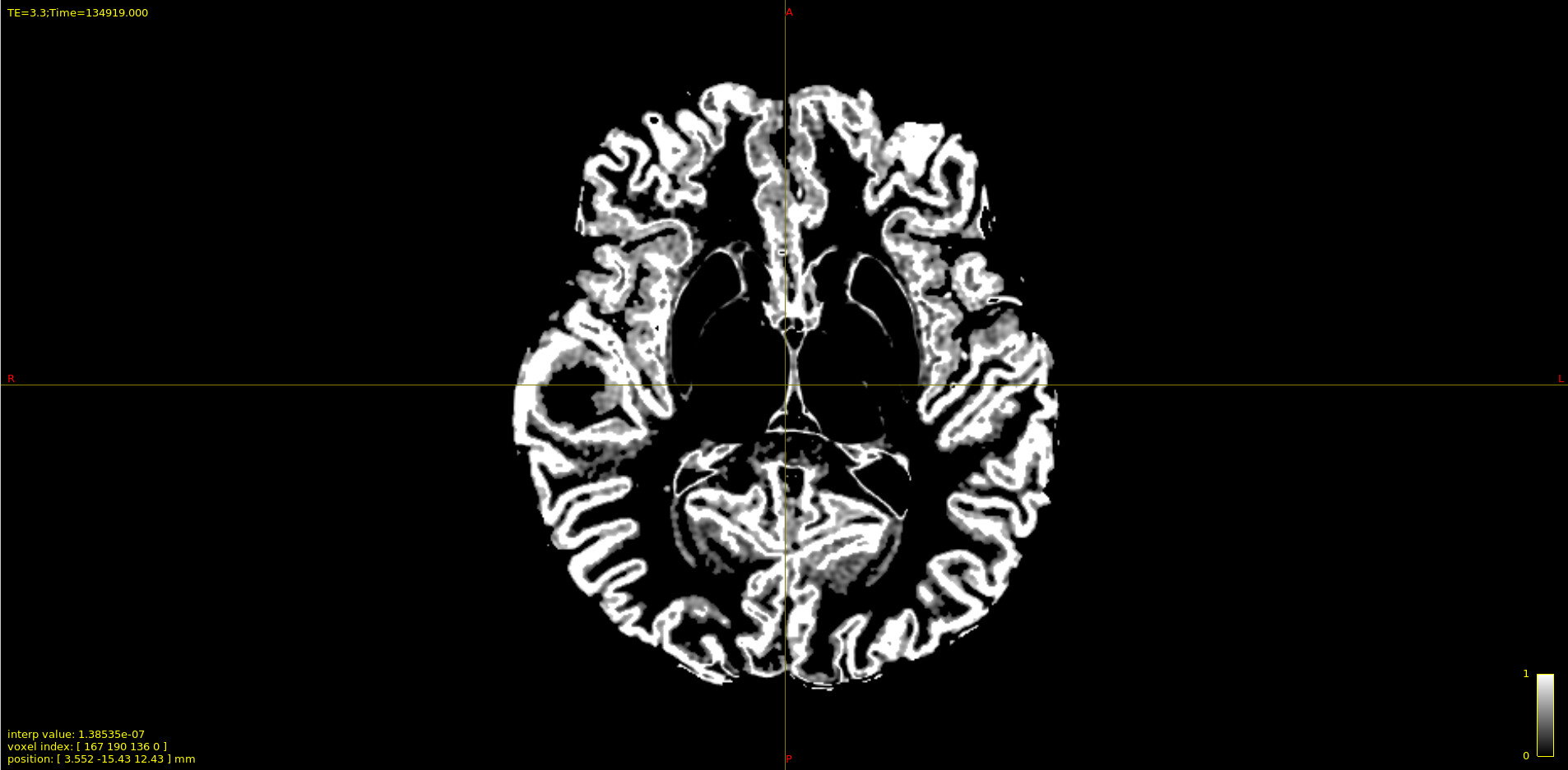
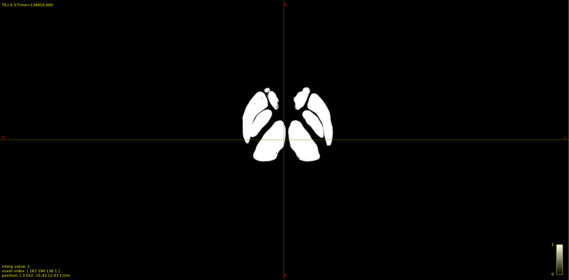
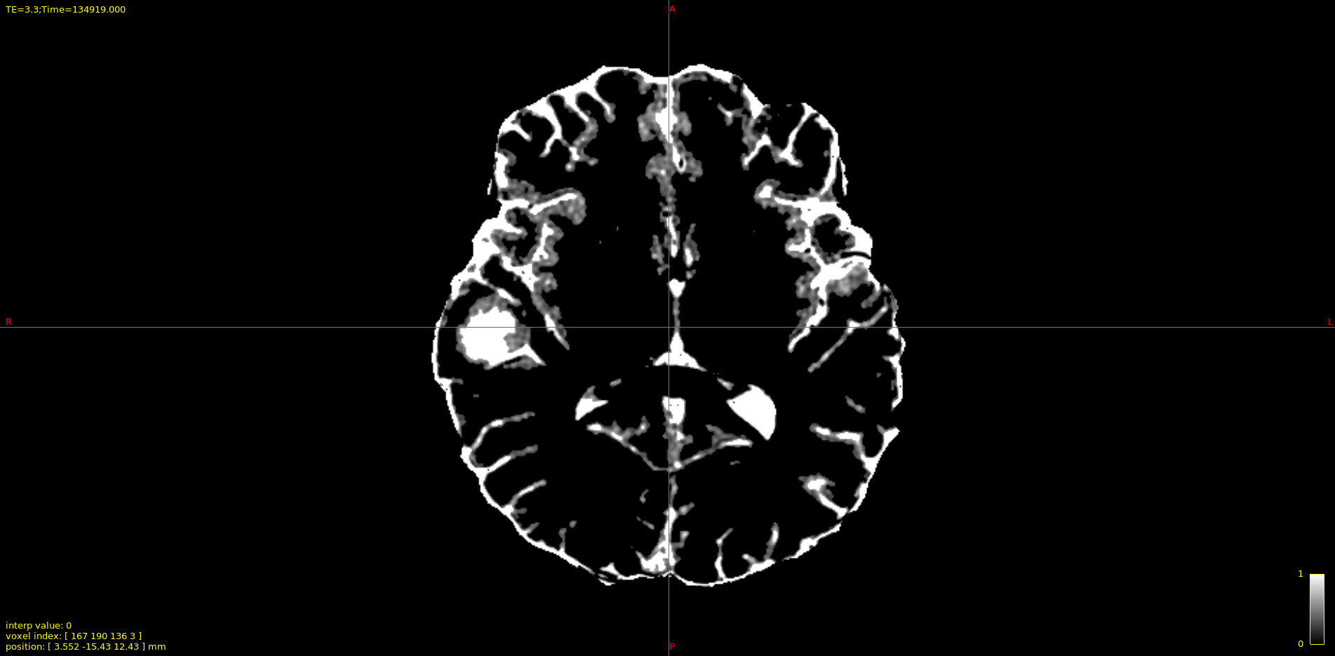
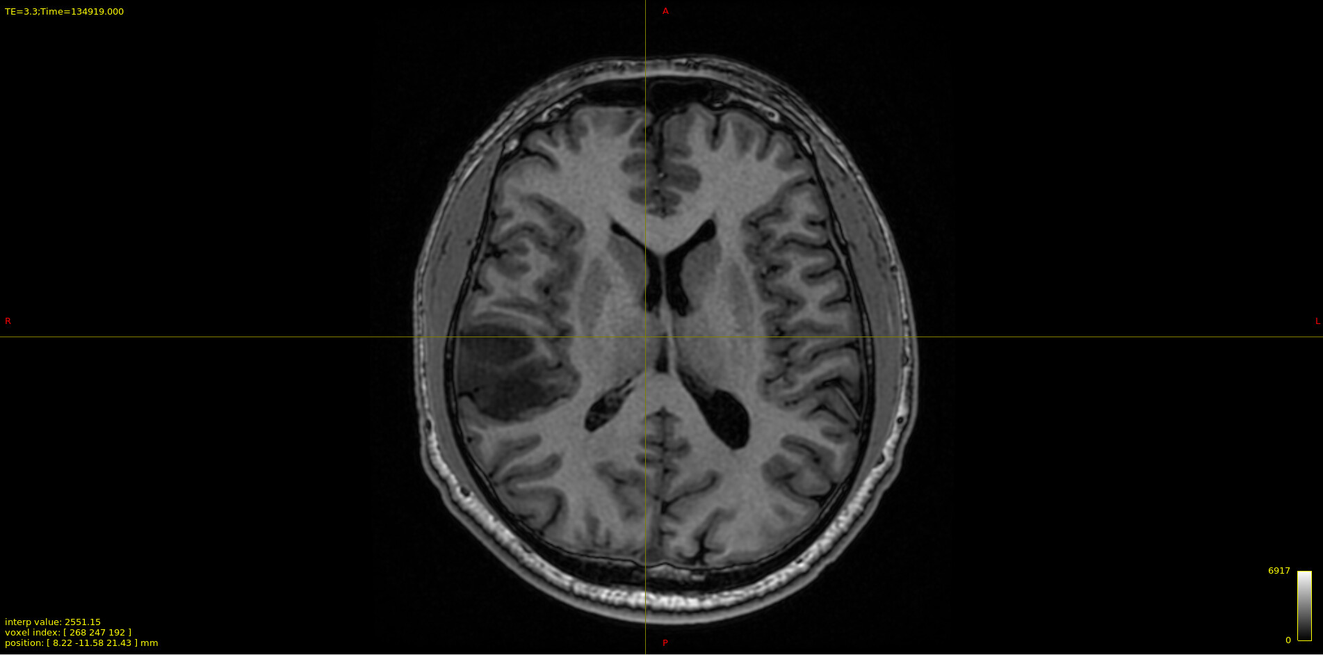
The results of the 5tt_nocoreg.mif file that I created with the 5ttgen function are as follows, but there is an issue. My original anatomical T1 images belong to a glioma patient, and our study is related to brain tumors. As you can see in the results, all tumor tissue that the 5ttgen function should pathologically detect has been included in the CSF portion. This is a problem because I believe it will cause issues when performing tractography for the connectome since the streamlines are formed to go from gray matter to gray matter.
Do you have any suggestions regarding this issue? In my opinion It is a very important problem for the study. Is it possible for me to manually segment the pathological tissue? We believe this faulty segmentation will affect the results of the study.
It may not actually be a problem… it depends on whether it is fair to assume that the underlying white matter fibres traverse the tumour or not.
Assuming that WM fibres do enter the tumour, then it would be fair to label the tumour as PATH (thereby permitting tractography streamlines to propagate into the tumour and be retained). Here are a couple of options…
manually segment the tumour (to create tumour_mask.nii.gz) and do something like 5ttedit <5tt_image> -path tumour_mask.nii.gz <5tt_image_modified>. This will result in a 5TT image that has the tumour as the 5th tissue type.
try a different 5TT algorithm. If FreeSurfer (or FastSurfer) can correctly segment the pathology, it could be worth trying something like 5TTgen hsvs <freesurfer_dir> <5TTimage_freesurfer> . It might be necessary to do another 5ttedit here. This also requires FreeSurfer or FastSurfer to be computed
Conversely, it could be possible that the tumour merely takes up space (and that WM fibres do not traverse the tumour). In this case, it would make sense to leave the tumour as CSF (this way, if a streamline propagates into the tumour area, it is discarded). The resulting tractogram will contain streamlines that do not traverse the tumour.
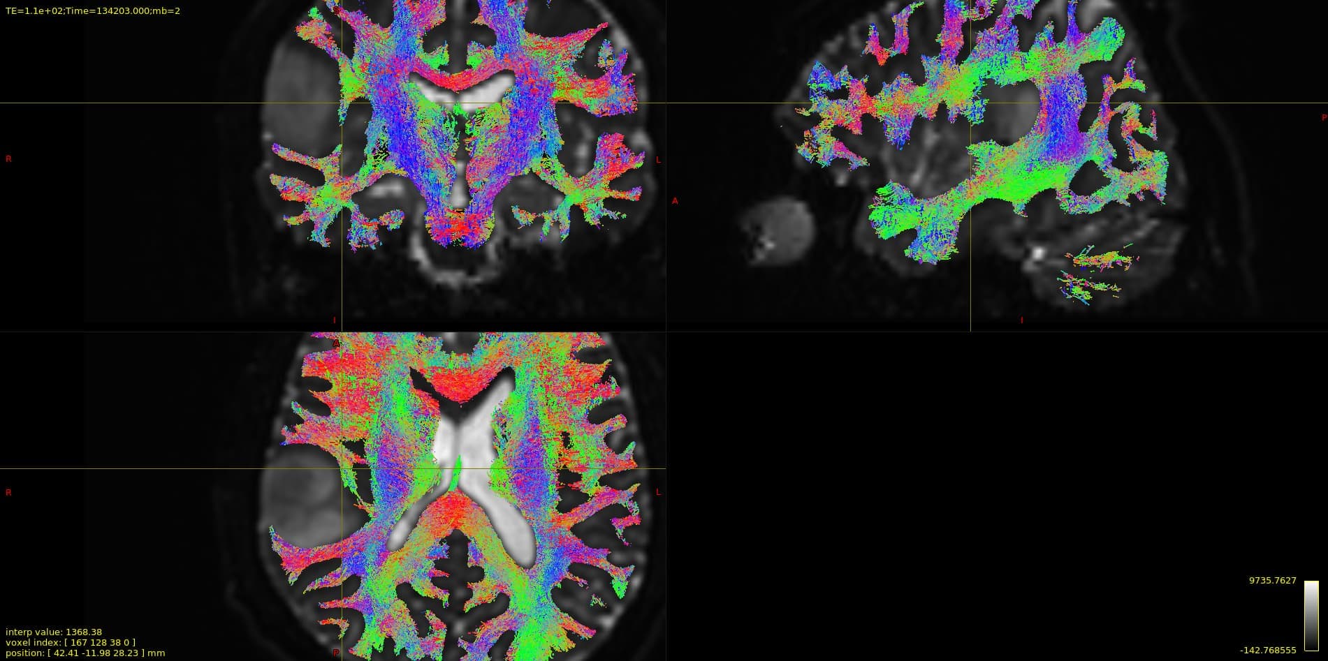
If you examine the tracts, you’ll notice that the streamlines seem to wrap around the tumor tissue, but logically, since there is gray matter around the tumor tissue, it would make more sense for the streamlines to be perpendicular to the tumor tissue. As you can see, this problem arises from 5ttgen mistaking pathological tissue for CSF when segmenting. (Because of that streamlines are passing from de side of the tumor in a parallel axis)
I will try your methods and get back to you as soon as possible!