I am a newbie to tractography analysis, trying to perform CSD and probabilistic tractography for the optic nerve (ON), with images of a single-shell DWI (b0, b1000, ~32 directions), and an additional inversed phase encoding direction T2 image and a T1.
I have preprocessed DWI images with dwidenoise, mrdegibbs, dwifslpreproc and registerred dwi to T1. Before tractography, I made the eyeball and optic chiasm ROI images based on T1, and a mask including the orbital cavity. However, after trying different algorithms in dwi2response (including dhollander, manual, msmt_5tt, tax, tournier) and estimating FODs using the txt file, the command tckgen iFOD2 could not generate ONs track between the ROIs with the generated FOD files, but tckgen Tensor_Prob with DWI worked and did a barely satisfactory job. It seems that the above dwi2response algorithms separate wm, gm and csf, but exclude ONs.
My question is here: is there any command in Mrtrix3 could perform CSD and probabilistic tractography on the ONs?
It shouldn’t be a problem, especially if you can obtain streamlines using the tensor approach. Could you provide more detail on the exact set of commands that you used? I think it might also be useful to show some screenshots of the ROI images that you used, and also the contents of the response files generated by the various commands you tried (it’s probably enough just to show those generated using the dhollander and tournier approaches).
Thank you very much for replying and offering to help.
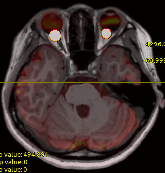
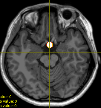
‘align.mif’ is the preprocessed file after registration. Below are some screenshots of the ‘R_eye.nii’, ‘L_eye.nii’ and ‘OC.nii’ ROI images of optic nerve heads and optic chiasm, respectively, and also the ‘align.mif’ overlay.
Below is the set of commands I used for CSD and FOD:
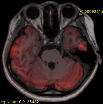
OK, given your WM FOD overlay image in that last screenshot, it looks like there isn’t much (if any) FOD intensity in the optic nerve – at least as far as I can tell? Can you display just the WM FOD image (specifically, its first volume) at the level of the optic radiations? And maybe also double-check that your new_mask.nii does contain the relevant structures?
Thank you very much for your help!
The new_mask.nii was checked to include optic nerves.
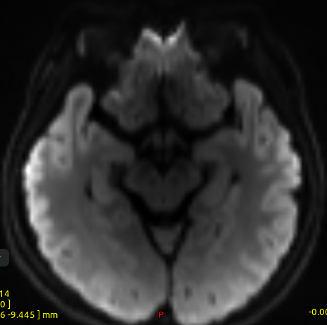
Here is the screenshot of the wmfod.mif at the level of the optic nerves:
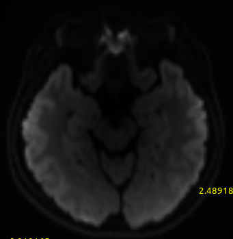
and the level of optic chiasm:
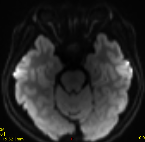
Moreover, I found that there is a small gap with relatively low FOD intensity at the level between optic nerve and optic chiasm like below, maybe it is where the nerves passing through the optic canal?:
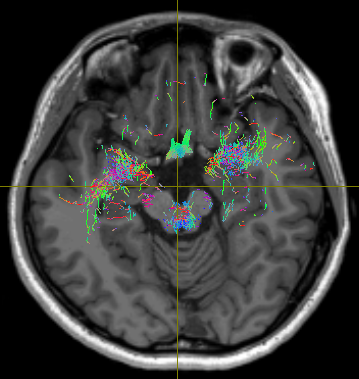
so I further tracked fibers with the ‘OC.nii’ ROI image with the command: tckgen -algo iFOD2 -cutoff 0.05 -angle 90 -maxlength 120 -seed_image OC.nii -seed_unidirectional -stop wmfod.mif fibs_OT.tck -force
it seems that the optic nerves were cut at that level before approaching the optic chiasm:
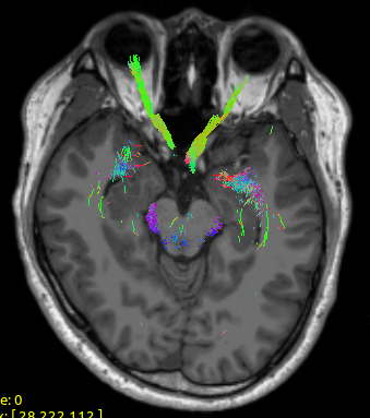
However, a few participants’ images (acquired with the sequence and analyzed with the pipeline) did not have this problem:
So maybe, is there anything I could do with the options or parameters of the dwi2response , dwi2fod and tckgen commands to enhance the FOD intensity at the optic nerve, or this is just the imaging problem.
Thank you for your help, again!