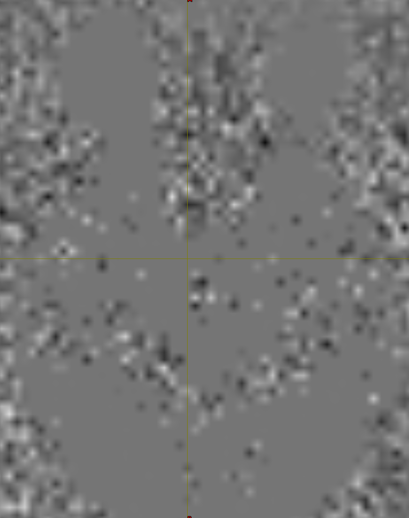
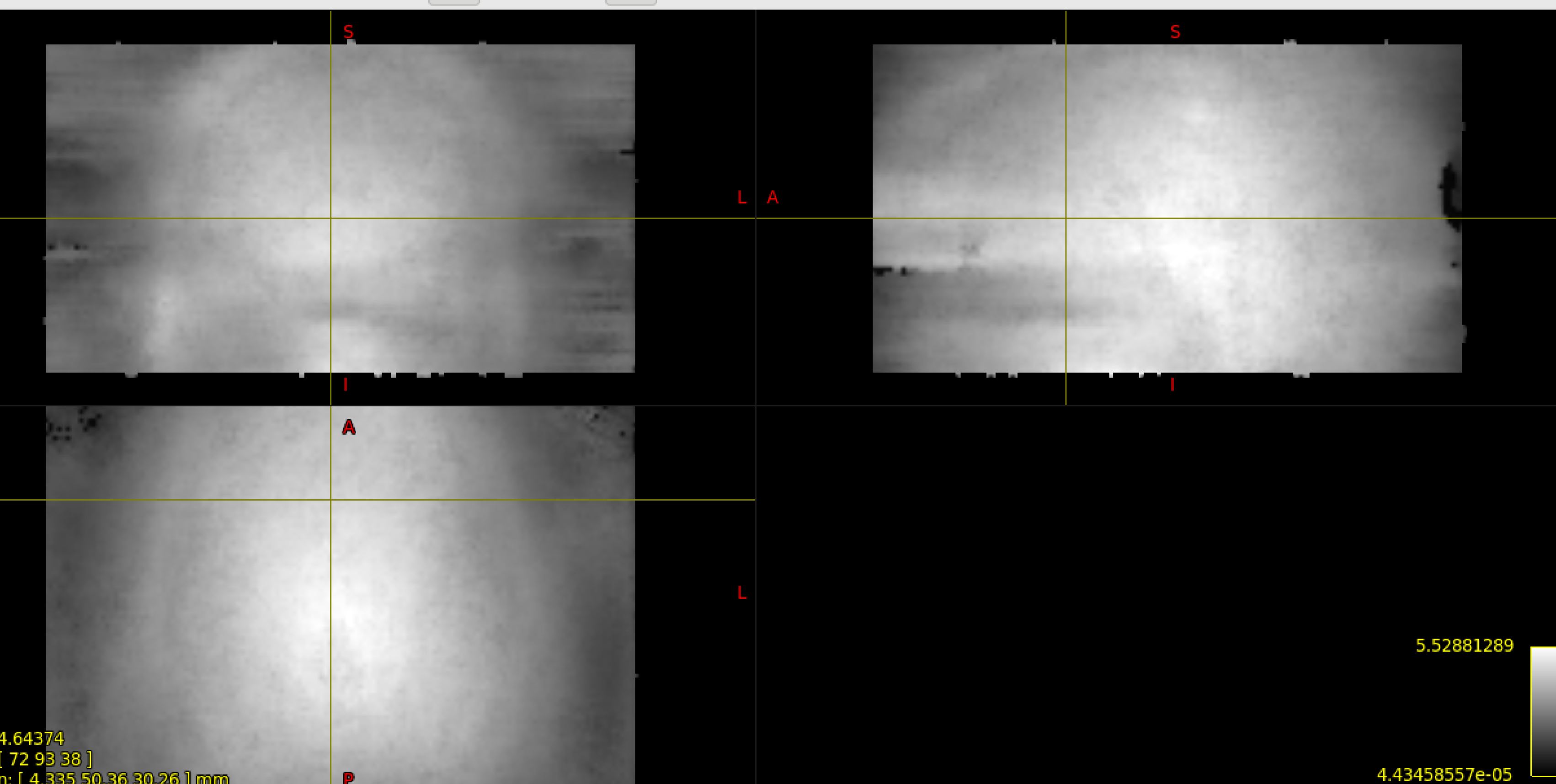
I had a question about my dwidenoise output. Reading the tutorials one expects not to see anatomical infomation in the residual maps.
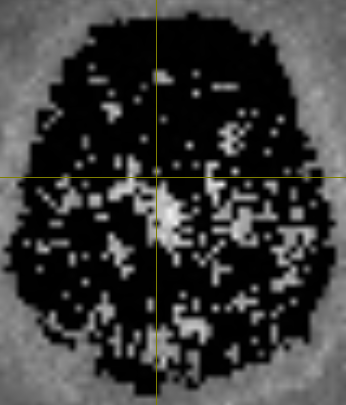
However in my residual maps I think I can see a bit of lateral ventricles (image 1) and an outline of the posterior fossa in image 2. Could I ask if these screenshots of my residuals look reasonable (appreciating its only 2 slices)? Thanks for your help as always.
That would seem to suggest that the denoising is not actually removing any (or very little) noise from brain regions. Maybe your acquisition does not have enough redundancy in it… Could you tell us a bit more about it (number of volumes, b-values, etc)?
However, maybe I’ve got the wrong end of the stick. Are you showing the noise map (as produced by dwidenoise -noise), or the residuals as suggested in the docs? If the former, then what you showed is very unexpected. However, if your image is a snapshot of the residuals, and this corresponds to the first volume, it might be that there’s not a lot of denoising going on for the b=0 (which is presumably the first volume?) - this wouldn’t be wholly unexpected particularly if you have a single b=0 volume. But the other volumes might still behave as expected. Have a look through all the volumes (use the left/right arrows), hopefully they’ll look more convincing. You could also compute an image of the RMS residuals across all volumes, which might be a better way to inspect the residuals in one image:
I was showing you the residuals as suggested in the docs. Looking at the other volumes there is a bit more denoising going on - (see example attached below). I have also attached an rms_residuals.mif which I presume looks as expected in the denoising? Is it also reasonable to assume that the fact there appears to be more denoising in the lateral ventricles (in the res.mif image) is because CSF is ‘nosier’ than neighboring white and gray matter?
OK, that still doesn’t look great… Looking at your acquisition parameters however, I note that you have 117 volumes, which is getting close to the 5×5×5=125 voxels included in the neighbourhood. We’ve noticed that there are issues with the denoising when these values get too close. The simplest fix at this point in time is to use a larger extent than the default, for instance a 7×7×7 patch size:
dwidenoise in.mif -extent 7 out.mif
In the meantime, @dchristiaens and I have been looking into ways to make the denoising more robust in these cases, but it might be a while before we produce a proper fix.
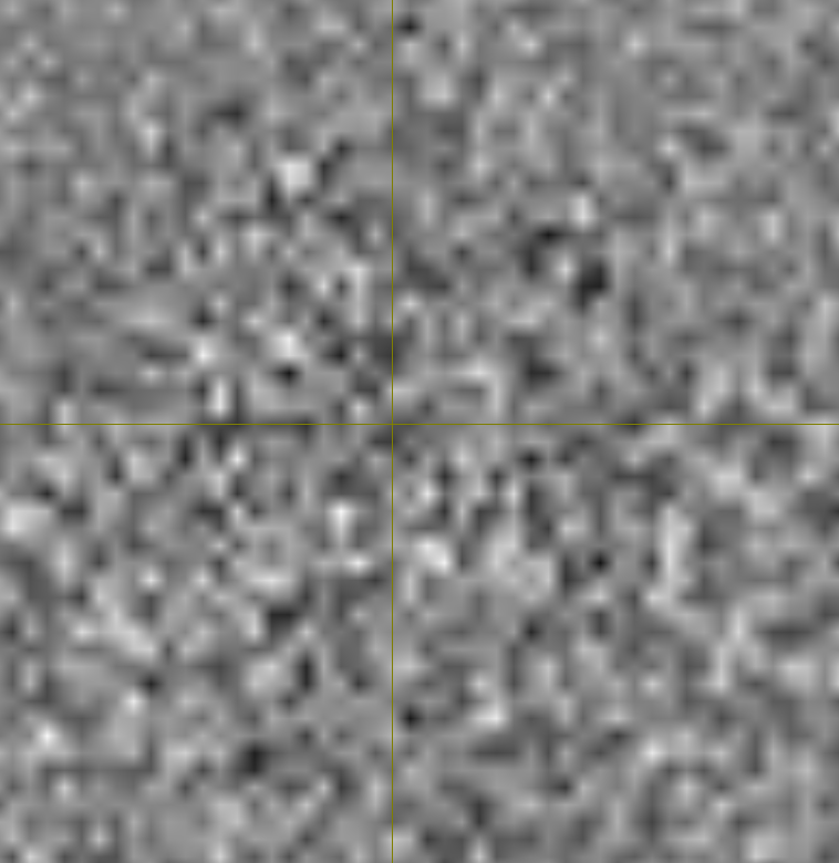
Thanks for that. Changing to 7x7x7 does completly change the look of the residuals now (see below). This seems to look more similar to the residuals as published in Verarrt’s paper though I still find it hard to detect a visable difference between the original and the denoised DWI.
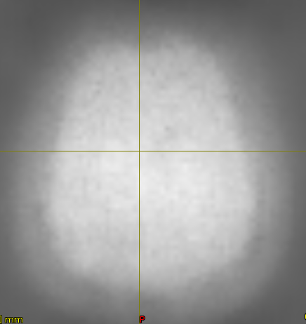
Yes, that definitely looks much better. Maybe also have a look at the noise map (dwidenoise output option -noise) to get an estimate of the SNR (average signal intensity, divided by average noise level).
I wanted to follow up on the last post, what appears to be the noise .mif file. For a multi-band, multi-shell acquisition, to what extent is seeing the structure of the brain (as shown above), bad?
I’ve seen EPI acquisitions that the noise looks more random, without much of the brain outlining along the GM visible. Is what is shown above bad? For our subjects we see a similar pattern, and those that use MRtrix in the group here are not quite sure if it’s a cause for alarm or not.