Hi, I’m a medical student in Brussels that helps out with the analysis of neonate DTI for a paper.
The paper researches wether the interhemispheric connections of the visual cortices is different between non strabism neonates (control) and strabism neonates.
In normal neonates, we expect a symmetry between tracts that start left and end right, and tracts that start right and end left. In strabism neonates, we expect a loss of this symmetry where tracts that start in one hemisphere, don’t end in a similar location in the other hemisphere.
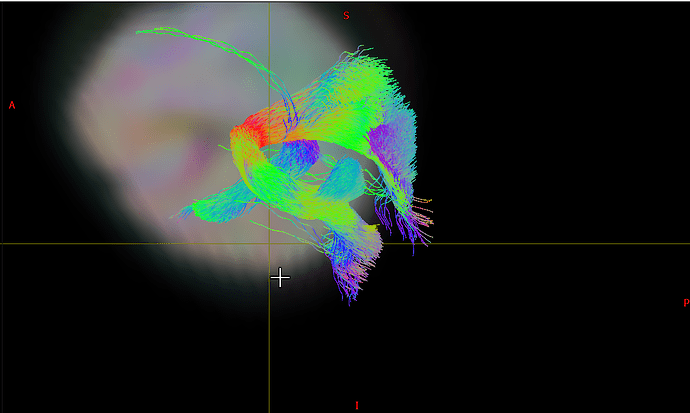
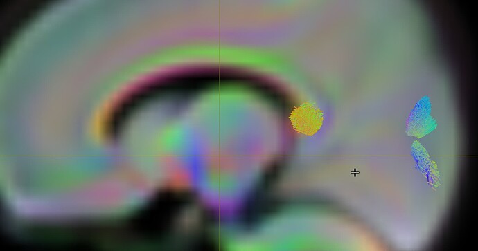
My trouble is that our tracts do not seem to start or end in the fissure calcarine, from wich we’re trying to extract the relevant tracts.
I need somebody to confirm the location of the fissure calcarine on the neonate atlas that we’ve been using, so we know that we can trust our tracts. My colleague is a physicist and thus cannot confirm the anatomical correctness of the images.
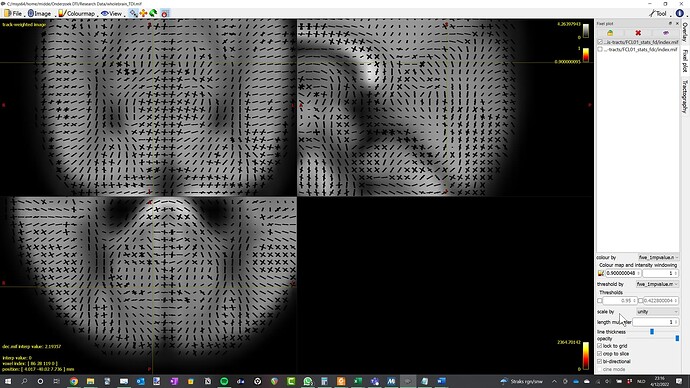
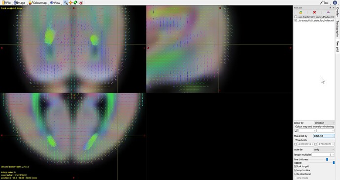
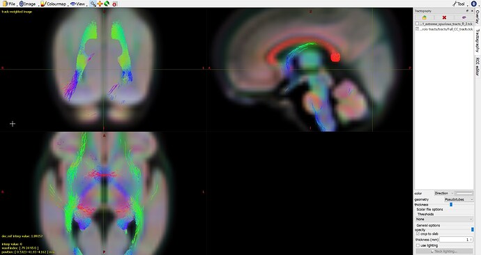
I’ve attached images of our atlas and the tractography that we’ve been working with. Below are also the methods that we’ve used so far.
Used Methods
The atlas has been generated based on 100 neonates, of which 4 had strabism.
To generate the tracts we’ve used 2 inclusion ROIs: 1 in the corpus callosum, 1 in one of the hemispheres.
To narrow the amount of tracts down, we used an exclusion ROI on the rest of the brain, so only tracts in the occipital lobe would be included.
A third inclusion ROI was used in the contralateral hemisphere to force Mrtrix to only include tracts that would specifically go down towards what we thought was the fissure calcarine. This yielded less results.
I hope someone could help me. If you need any more data, tracts or ROIs, I’ll be glad to help.
