Hi,
I am going to extract Corpus Callosum fibers. After following the initial steps, I used “tckgen” command and chose my saved ROI (which was segmented by fsl) as a seed_image :
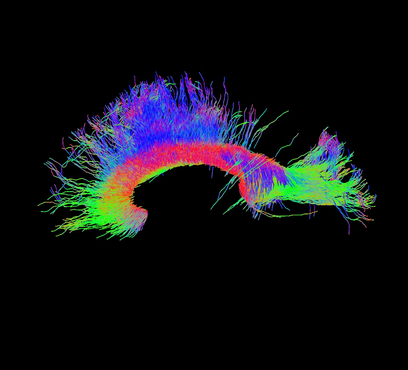
The problem is the extracted fibers are not complete. You can see the result in the attached file. I check my ROI and fiber orientation distribution file. They look both alright. I have used this method for multiple subjects and a few of them had this issue, most of them were alright.
When conducting CC tractography, it is often important to make the mask (from sagittal) more than just 1 (mid-sagittal) slice. That is, I usually do 3 sagittal slices: 1 from mid-sagittal, and then 1 from the both sides as well. If you only do 1 slice (mid-sagittal), then you usually get everything but the isthmus (which I think is what happened in the example you uploaded). Sometimes you will also get crossing fibres (which extend inferiorly in the corticospinal tract; CST) so you may need to place small exclusion masks to ensure they are not tracked. You will know when you have tracked the CC properly as you will also see the tapetum (1 on each side).
And/or try reducing the FA threshold (i.e. reducing the -cutoff value).
Hi Jerome,
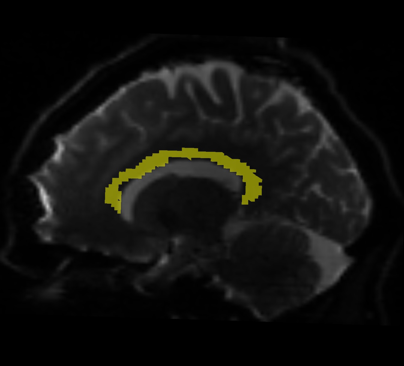
Thanks for your reply. I segmented the whole corpus callosum using the standard mask by FSL. So it is not just one mid-sagittal slice. I tried changing the cutoff value and also the number of fibers, then I got a better result. But the point is because I want to extract CC for multiple subjects and do some statistics test on them, I wanted all the parameters for extracting the fibers to be the same. I do not know how much it is important and if I set different cutoff values, does it affect my final result or not. Because for the 10 subjects I’ve done so far, just 2 had this problem and others showed all CC fibers properly.
It’s hard to diagnose from screenshots alone, but there are a lot of experiments that you can do with the data to try to gain insight here. For instance, manually draw a ROI in the volume where you think those streamlines should be going, seed streamlines there, and see where they go. Also draw a ROI in the CC with a reduced volume corresponding to the part of the cross-section of the CC where you think those streamlines should be, and repeat the tracking experiment seeding from there but with different cutoff values. With higher cutoff values, streamlines are more likely to stay “latched” to the most collinear FOD lobe rather than bend and start following another. Also check to see if there are streamlines that are trying to curve in that direction but are terminating prematurely.
If you find that that particular pathway is better reconstructed seeding from the cortex and tracking toward the CC rather than the other way around, you could try performing whole-brain tracking and then selecting the streamlines corresponding to the CC after the fact.
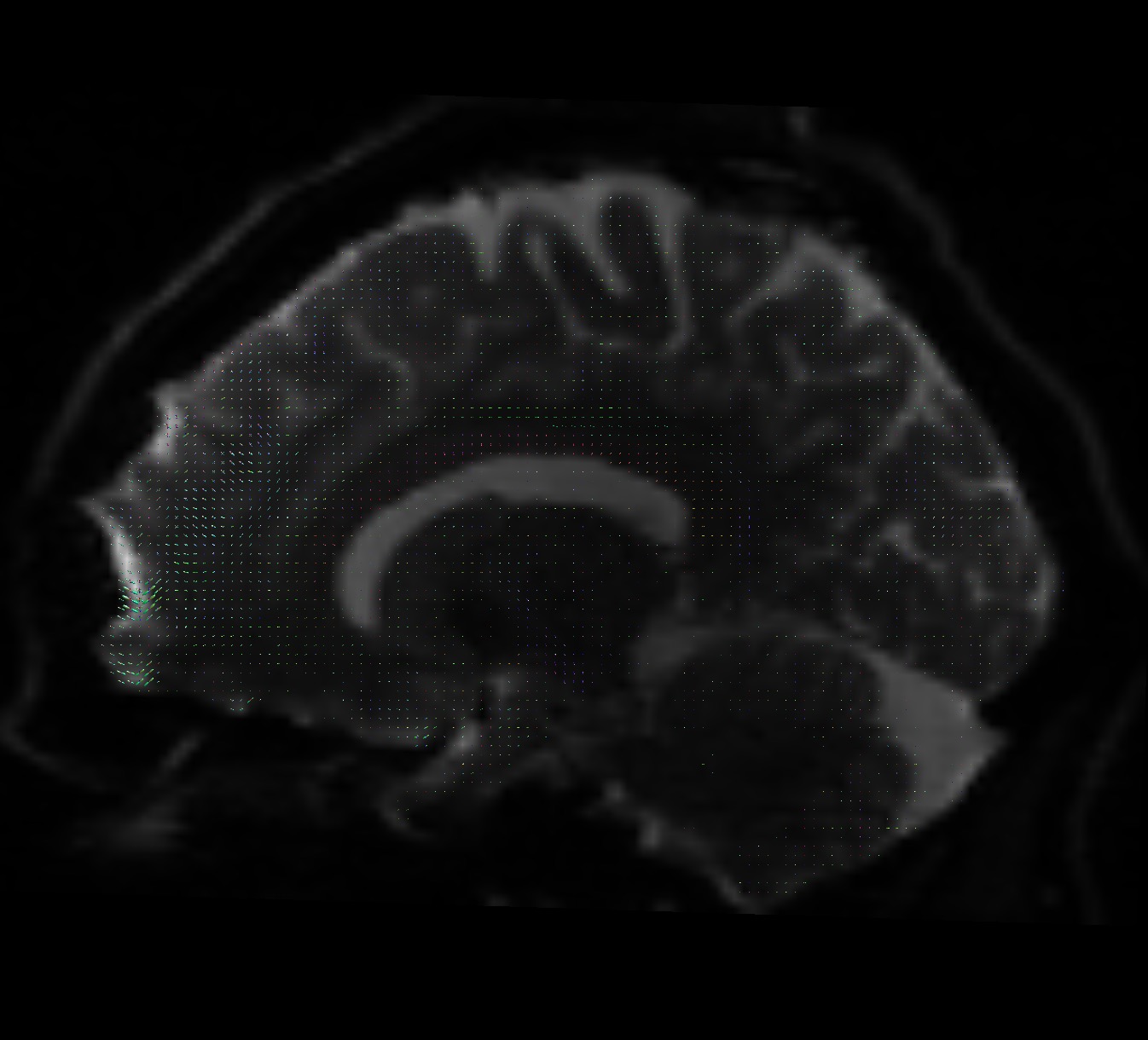
From the perspective of checking the data, what would be of most use would be a coronal slice within the plane where the streamlines are going only laterally and not medially, showing the FODs.