I am writing to seek advice regarding a tractography issue encountered during my research on ex-vivo mouse brain dMRI data.
Data and Modeling Overview Data Acquisition: I am utilizing multi-shell data with b-values of 1000, 2000, 4000, and 6000, totaling 16, 20, 30, and 40 directions, respectively, alongside four b0 volumes. Preprocessing: Data has undergone denoising, degibbsing, eddy current/motion correction, and bias field correction. Modeling: I used the dhollander response function estimation followed by Multi-Shell Multi-Tissue CSD to generate the White Matter Fiber Orientation Distribution function wmfod. The wmfod image accurately distinguishes between white matter and gray matter.
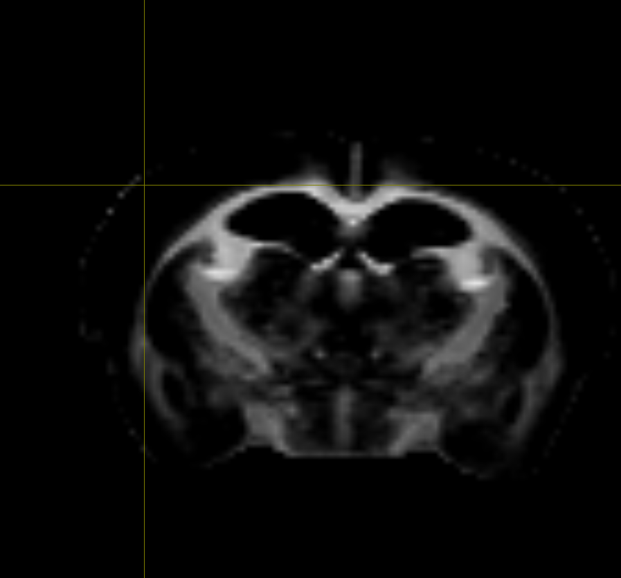
The problem is missing cortical tracks
My primary goal is to visualize and quantify crossing fibers in the cortex region.However, when I use the wmfod file as the input for tractography (e.g., using tckgen), the streamlines consistently terminate or are heavily suppressed upon reaching the Gray Matter. Consequently, I obtain almost zero tracks in the cortex, which prevents me from studying the local cortical fiber architecture.I wish to leverage the higher accuracy provided by MSMT-CSD results for this analysis.
Could the community please offer guidance on how to resolve this typical issue?
Tractography Method: How can I use the wmfod from MSMT-CSD to successfully generate streamlines that enter, traverse, and terminate within the cortex?
Parameters/Techniques: Should I drastically lower the FOD amplitude cutoff value used in the tracking algorithm? Or are there specific techniques (such as implementing ACT or utilizing other FOD files generated by MSMT-CSD) that are required to achieve tracking within the low-signal GM areas?Any practical advice on resolving this MSMT-CSD/Gray Matter interface challenge for ex-vivo data would be greatly appreciated.
Firstly, utilising ACT naively has the prospect of completely forbidding the tracts that are of interest to you. If you were to explicitly segment the cortex and place it in the first image volume, then ACT would impose the prior designed for cortical grey matter in adult human data, which is “don’t enter me”. So that would totally contra-indicate your intended outcome. One approach that I’ve suggested in a few contexts is to send that tissue parcellation to the second image volume, which is intended as “sub-cortical grey matter for adult human data”, and will permit streamlines to continue propagating within it, all it will do is forbid a streamline from going from the white matter into the cortex and back out again. So would probably have minimal impact on your pursuit; and indeed would preclude reconstruction of streamlines exclusively within the cortex, since currently the ACT implementation forbids streamlines from existing only within sub-cortical GM and never entering the WM.
Firstly, I would stop treating this as a tractography problem and start treating it as an imaging problem. Attempting to diagnose or rectify problems based on tractography is the wrong premise if the tracts are not visible in the FODs to begin with.
The first experiment I would suggest is running single-tissue CSD with single-tissue data. The question you need an answer to is: are these tracts simply not visible in the DWI signal, or are they present in the signal but specifically the MSMT-CSD modelling approach is not recovering them? Yes doing single-tissue CSD might result in FODs that are noisier and less specific, but it might contribute to answering the question of whether there’s any signal there at all.
Now for the fundamentals. What will crossing fibres in the cortex look like, given MSMT is given the task of disentangling the contributions of different macroscopic tissues to the empirical DWI signal.? Well, maybe it looks like a linear combination of one component that has a decay curve that looks like the “GM-like” response function and is isotropic, and a second component that has a decay curve that looks like the “WM-like” response function and is anisotropic. In this scenario, assuming your data are of adequate quality, hopefully MSMT will do reasonably well. But maybe those cortical crossing fibres have a decay curve that looks like the “GM-like” response function but are anisotropic. What will MSMT do in this instance? Well, it can only try to find a suitable linear combination of the “GM-like” and “WM-like” response functions that best match the empirical data. So maybe it ascribes a bit of signal to the GM-like component and a bit to the WM-like component. But how much to each? Well, it will depend on your acquired data and the discrepancy between the response functions. One factor here I’ve been concerned about for some time is that inclusion of the intermediate b-values, where WM and GM are comparable in magnitude, can reduce the separability of the problem, and perhaps kind of “smudge” signal between those two ODFs. But I digress. I think that if you spend enough time on the problem, you might come to the same realisation I have, which is that there is a fundamental conflict between attempting to resolve crossing fibres in the cortex, and doing a deconvolution with an isotropic GM-like kernel and hoping that some of your cortex instead gets labelled as “WM”. Yet another project I wish I had the resources for…