Hi, I wondered if there is a way to import TMS data as ROI with a segmentation tool. Until now I was not able. I have the TMS points as DICOM and can import the data set to MRtrix, drawing the ROIs manually would be a lot of work…
As I mentioned on ResearchGate, I don’t have much experience with TMS myself. Maybe to help us understand your problem: what kind of data do you get from TMS? You say it’s in DICOM: is it an image you can open in MRtrix? Can you show us an image / screenshot? If so, stringing together a couple of commands in MRtrix could turn it into ROIs, depending on what you’re after…
Hi, yes, we work with TMS data.
The TMS-Spots, where you have a motor, speech or whatever reaction, depending on what your interest of stimulation is, are burned-in or based on the T1 mprage. You can export both the negative and positive spots and you can decide if you need the anatomical info or not – so you export the spots based on the T1 with or without anatomical info. If without, you have an image with 192 or so slices (T1) and the markers (nose & ears) and white spots (positive voxels) in black space. If with anatomical info, you have the structural T1 plus burned-in white spots.
For tractography, I want usually control and detailed info, so I draw single ROIs for each TMS spot and see, what seeding from there results in.
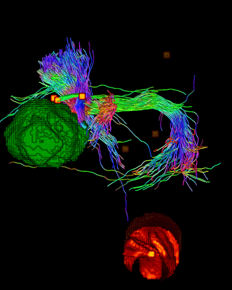
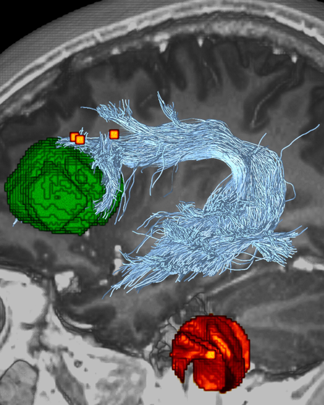
I attached two images where you can see positive TMS spots for speech in relation to tumour segmentation (via itk-snap) and fiber bundle of interest (AF). One image with seeding from one of the spots (just the TMS-positive spots image – you can see as well the darker contralateral spots) and one without seeding from the spots (with the T1 and overlayed TMS spots). At the bottom, you can see the marker for the left ear (circle shaped).