Hi MRtrix3 team and community,
I am seeking advice regarding an unexpected finding during a Fixel-Based Analysis (FBA) comparing two cohorts of healthy controls.
Context & Data
We compared two independent cohorts of healthy controls acquired with slightly different scanning timeframes/protocols on the same general setup:
-
Control Group A: 24 healthy subjects
-
Control Group B: 14 healthy subjects
-
DWI Acquisition: Imaging was performed using a 3.0-Tesla MRI scanner (Ingenia, Philips) with a single-shot EPI sequence at a b-value of 1000 s/mm² (128 diffusion directions). Single-shell b= 1000 s/mm2 with 128 diffusion directions. (Preprocessed with standard denoising, degibbs, eddy/motion correction, and bias field correction).
-
FBA Pipeline: Standard Recommended Pipeline (single-tissue CSD for single-shell data, population template generation, non-linear registration, fixel cross-section and density computation, statistical inference via fixelcfestats with CFE and 5,000 permutations, covarying for age and sex).
Phenomenon
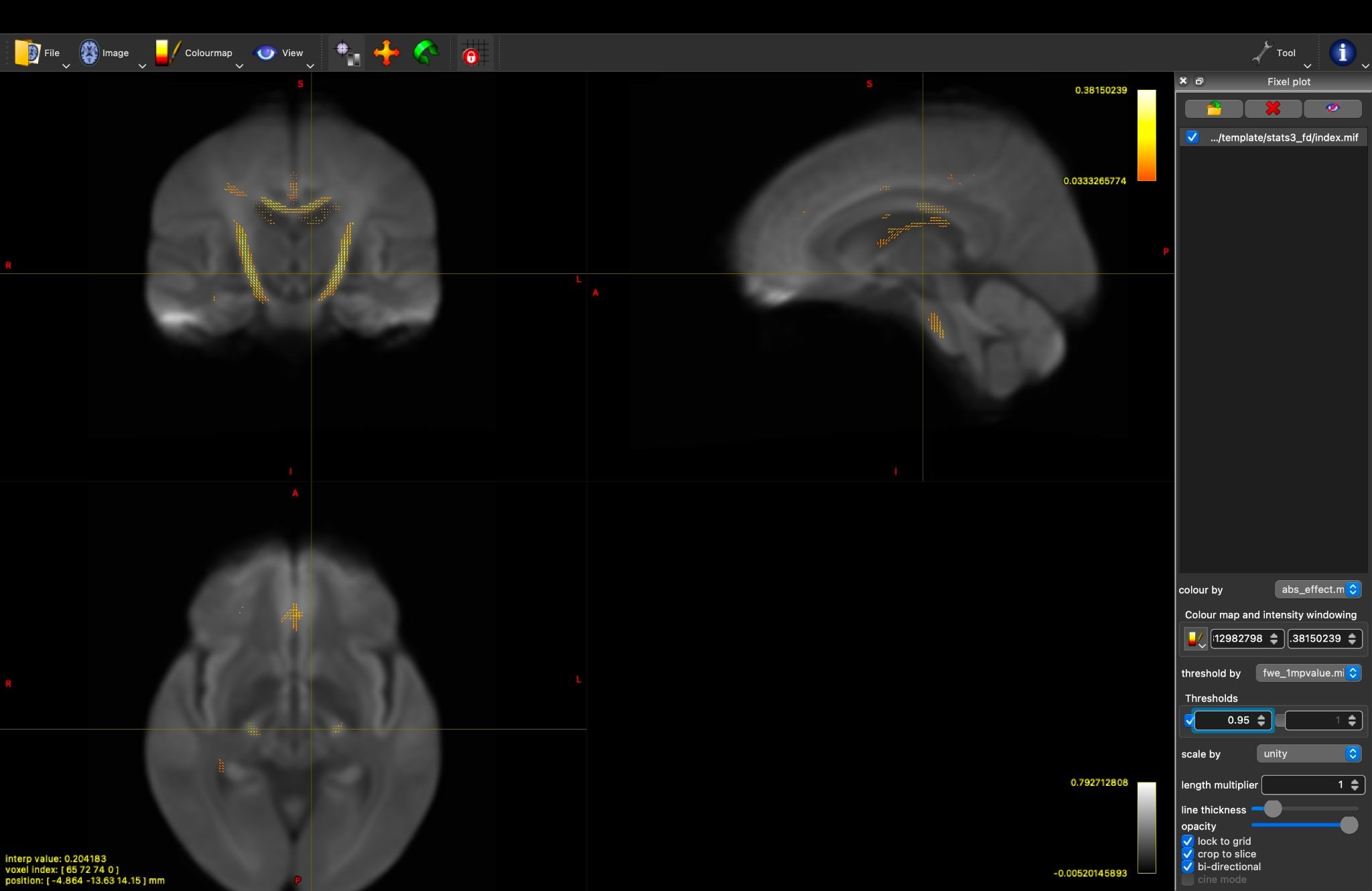
Despite comparing two healthy control cohorts (where we expected no significant structural differences), we observed significant group differences (pFWE < 0.05) specifically along the corticospinal tract (CST) across FD, FC, and FDC (Figure 1, 2, and 3). Rather than a global, diffuse baseline shift across the whole white matter, the “batch/protocol effect” appeared concentrated almost exclusively in the CST.
Figure 1. FDC Figure 2. log_FC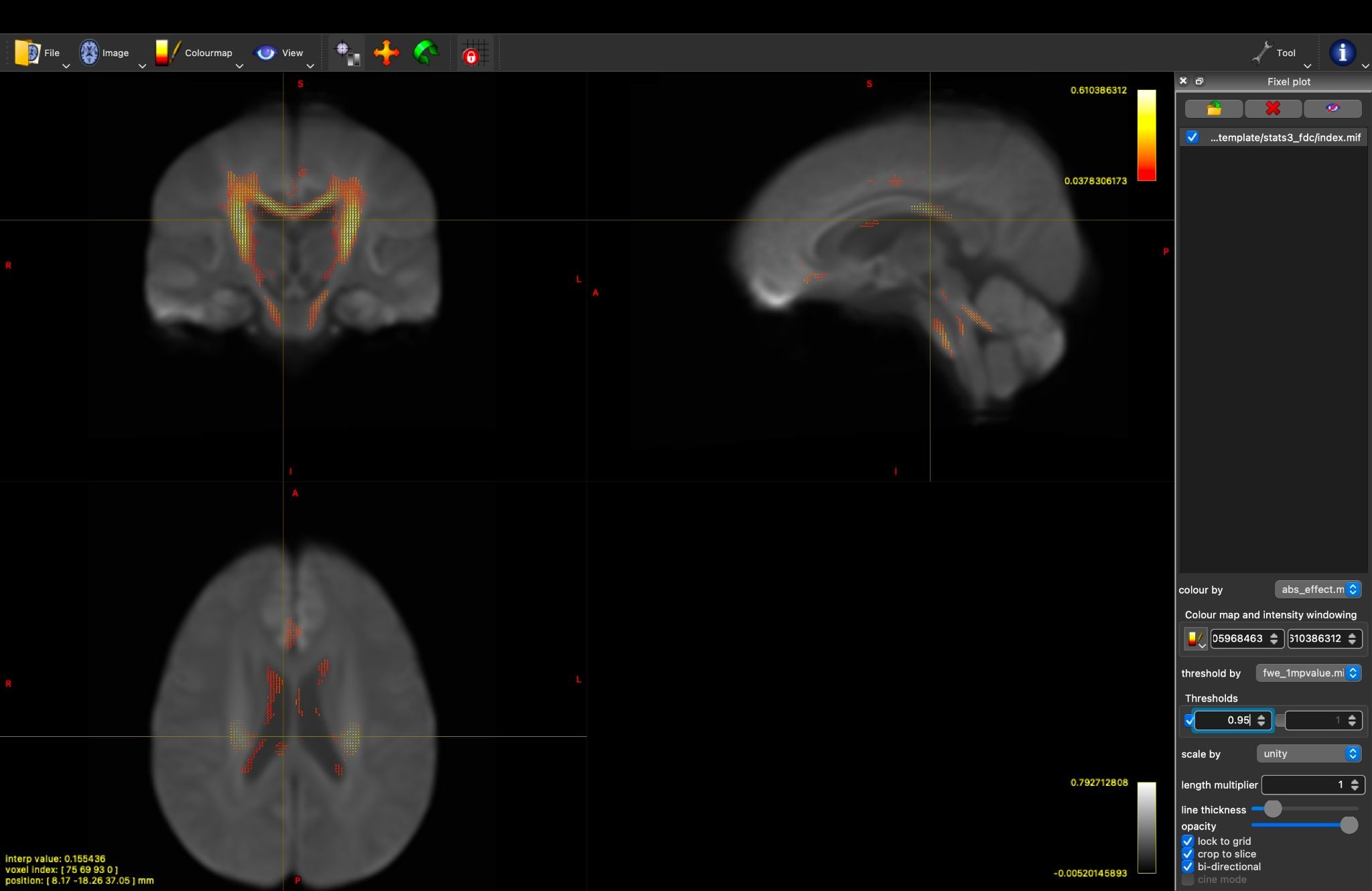
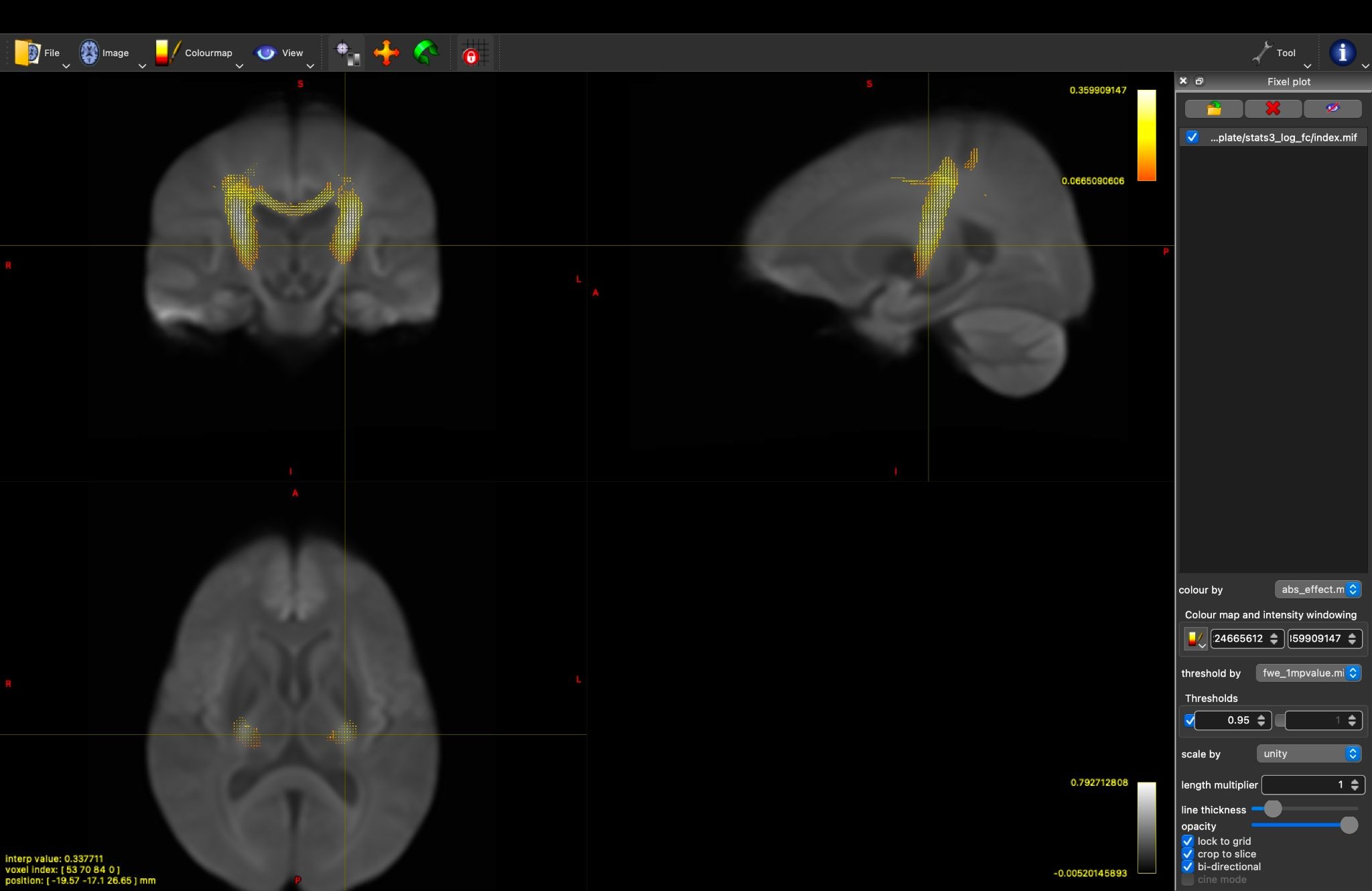
Figure 3. log_FD
Questions
We understand that MRtrix3 strongly recommends multi-shell or higher b-values (b > 2500 s/mm2, directions >45) for optimal CSD resolution and elimination of isotropic free water, and that single-shell b=1000 data has known constraints.
Given this context:
-
Biological vs. Algorithmic Localization: Why would scanner/protocol drift or batch effects manifest so selectively in the corticospinal tract rather than globally? Could the combination of high directional packing (single, prominent fiber population aligned along the z-axis) and gradient non-linearity/eddy currents make the CST particularly vulnerable?
-
CFE Enhancement: Could the Connectivity-based Fixel Enhancement (CFE) algorithm inadvertently amplify small, continuous systematic drifts along long, coherent tracts into statistically significant FWE-corrected clusters?
-
Handling Batch Effects in FBA: In the literature, ComBat is widely used for scalar DTI, but how does the community currently recommend harmonizing or controlling for subtle protocol differences when running FBA on fixel-wise metrics (FD/FC/FDC)?
Any insights, technical perspectives, or relevant literature pointers would be greatly appreciated.
Best regards,