I’m working on a tractography pipeline to extract optic radiation using MRtrix3. My standard script includes coregistration steps between DWI and T1 images using flirt and transformconvert, followed by applying the transformation with mrtransform.
However, for some of my subjects, the T1 and DWI images are already aligned in scanner space — same voxel size, orientation, and alignment. I can visually confirm this in mrview, and the headers match.
My questions:
If the T1 and DWI images are already aligned, is it safe (or recommended) to skip the registration step (i.e., no flirt, no transformconvert, no mrtransform)?
In this case, I wouldn’t have a diff2struct_mrtrix.txt matrix file. Should I simply use the untransformed 5tt and atlas masks as-is in DWI space?
For things like LGN or HCP-MMP1-based parcellation masks, is it reasonable to assume they are aligned too (assuming generated from the same subject and space)?
Thanks in advance! Any insights or caveats would be much appreciated.
if the T1 and DWI images are already aligned, is it safe (or recommended) to skip the registration step (i.e., no flirt , no transformconvert , no mrtransform)?
There should be no harm in including the registration step, even if the images are already aligned for some participants. In such cases, the transformation will essentially be an identity, so it won’t alter the data. The advantage of applying the same registration workflow to all participants is consistency: it ensures that any differences observed downstream are less likely to stem from differences in preprocessing.
If some participants go through coregistration while others do not, there’s a risk that group-level differences might reflect methodological inconsistencies rather than true anatomical or physiological variation.
In short, I’d recommend running the same pipeline for all participants. Subsequent steps (such as generating 5TT images, applying parcellations, and performing DWI analyses) can then proceed with confidence that all inputs are aligned in a consistent manner.
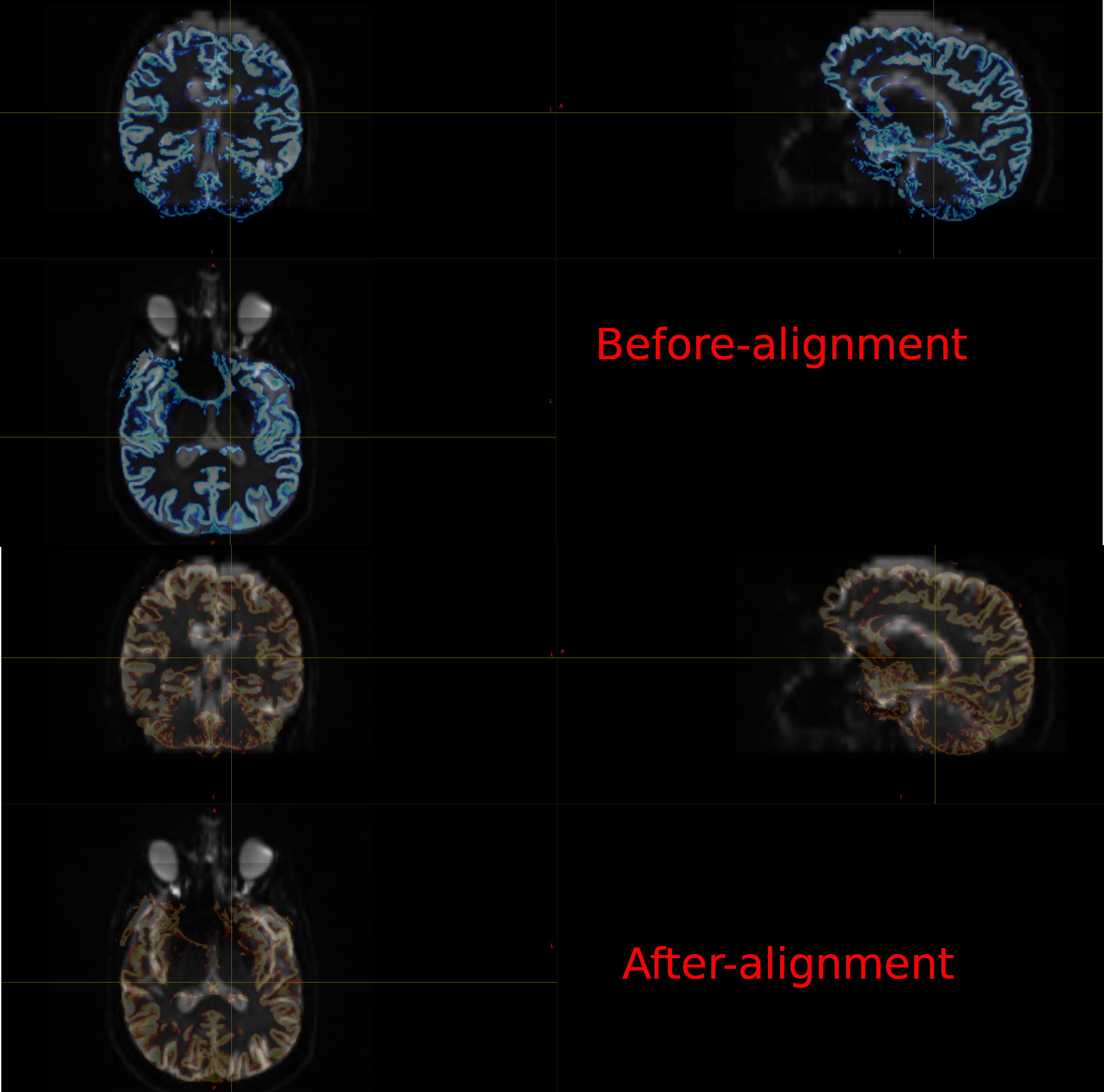
I am sure that you are aware of this, but based on the uncorrected DWI image in the above picture, make sure that you properly correct for any distortions/biases in the DWI (Gibbs ringing,bias/normalisation,topup/eddy, …)
Also, make sure that, for example, the T1 images you are using are not corrected for gradient distortions. Depending on your scanner vendor, scanning protocol, and setup, gradient distortion-corrected structural images can be output if desired.
Concerning, your point 3, if proper registration and corrections have been applied, your assumption might be reasonable. However, I recommend always checking to confirm this is the case!
Depending on the type of analysis you are planning, this might be a concern because it suggests you are mixing data sets either from different sources, or from different processing pipelines.
You’re right — all patients should be processed using the same standardized system, so transformation is indeed necessary. I’m currently working on optic radiation tractography. With the current method, the occipital and thalamic regions appear to be well-aligned; however, I still have some uncertainties.
From what I understand as I read through the methodology, these masks primarily serve as constraints or seed points for the tract orientation rather than rigid anatomical boundaries — is that correct? I’d appreciate it if you could clarify this point.
Also, would you recommend using EPI-based registration instead of FLIRT? I’ve come across several sources suggesting that epi_reg offers better performance, especially when aligning diffusion and anatomical images.
Additionally, I’ve read that incorporating a brain mask can improve the quality of results. What’s your recommendation regarding that?
What are the potential implications of using gradient distortion–corrected T1-weighted images?
This depends on how the masks have been defined in the 5TT image and the selected seeding mechanism in tckgen
@pyotrfrancois as for registration method, I’m a fan of epi_reg with the white-matter mask option (here’s an example that might be helpful). In saying that, I’ve seen flirt do a good job, too. It might be worth trying both approaches and visually inspecting the results.
@adsouza, if using at the scanner generated gradient distortion corrected T1 there might be addtioanal shape differences between the T1’s and typically-not-at-the-scanner-generated-gradient-distortion-corrected DWI images that might to be considered. I just brought it up as an hypothetical as the given information about the exact constellation of imaging data was sparse