This is probably due to the diffusion gradient table orientation being wrong - you can run dwigradcheck and correct this in your diffusion data before obtaining the FODs.
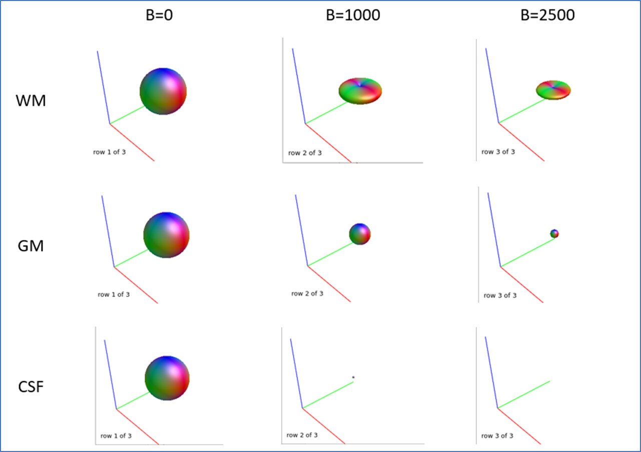
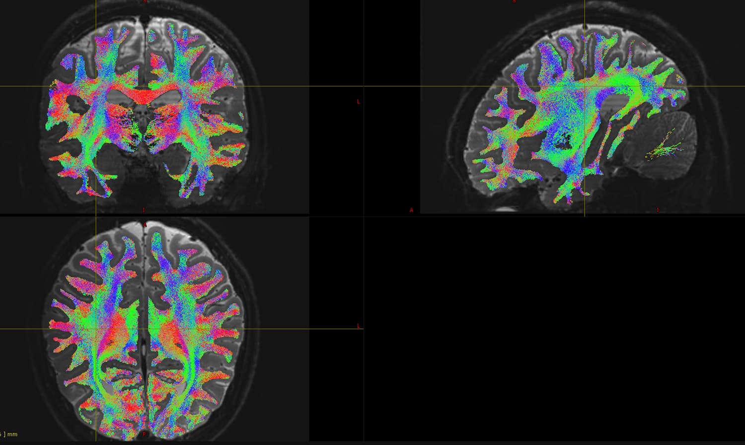
Actually, I don’t think there’s anything wrong with the directions of the streamlines (i.e. the local tangents). I suspect the issue is more likely to be that the acquisition is not of sufficient quality to resolve the multiple fibre crossings in the centrum semiovale, which the fibres from the corpus callosum would feed into. It’s not unusual to see streamlines heading back down the corticospinal tract like this (I assume this is what you’re concerned about?). This can happen especially when the angular resolution is poor, or the b-value too low, or the overall SNR isn’t high enough, etc.
Can you provide details about your acquisition protocol and the commands you used to process the data?