We’ve recently come across a problem with data acquired on a 3T Philips Ingenia CX, which seems to have occurred after upgrade from Philips software 5.6 to 5.7, which happened for us in August 2021.
The problem arose in a study on leg muscle architecture (muscle fibre orientations), where we started to notice a clear difference in fibre orientations after the upgrade, where primary eigenvectors were more aligned with the z-axis than they should be. Diffusion properties (MD, FA, primary eigenvalues) all seem fine, suggesting it has got something to do with the b-vectors. So we followed recommendations to validate DTI primary eigenvector orientations by obtaining scans of the same subject with various angulations (NITRC: dcm2nii: Document Manager: Display Document). We obtained four scans: one with the image axes aligned with the scanner, one with a 20 degr rotation applied along the right-left axis, one with 20 degr rotation around the anterior-posterior axis, and one with rotations applied in both planes (which is how we typically obtain our scans). The results were surprising, and confirmed there is a problem:
-
Even without image angulation, the primary eigenvector orientations seemed incorrect - this is based on our knowledge of leg muscle architecture and inconsistency of tractography reconstructions between scans obtained before and after the software upgrade.
-
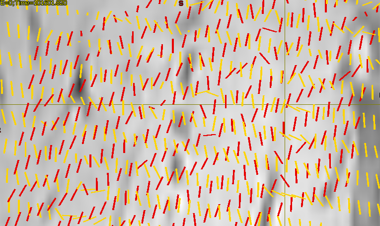
The angulated scans had fibre orientations that were different from the non-angulated scan, and different from each other. This was clear when visualizing the primary eigenvectors of all four scans using the fixel-plot option in MRview. (As they were all obtained during the same scanning session without the subject moving, the orientations should be consistent when viewed in scanner coordinates.) Below is a screenshot of a zoomed-in sagittal slice where the difference in fibre orientation between not-angulated scan (yellow) and AP-rotated scan (red) is clear.
We’ve posted about a related issue before (Tractography error with Philips scanner), but I hope this post will trigger responses from others who have had similar issues, particularly with Philips scanners after the upgrade to 5.7.
Also note that this is the same dataset we’ve previous had DICOM sorting issues with, which have been resolved in the latest MRtrix release: Problem with sorting of DICOM images using mrconvert
Any help would be appreciated. Also, I’m happy to share the data we used for validation if that’s helpful.
Cheers, Bart